
Ear Shaping
Parts of the Ear: A Parent Guide to Newborn Outer Ear Anatomy
Learn the parts of the outer ear—helix, antihelix, concha, lobule, tragus—and how newborn shape differences relate to ear molding.
Parents often search for the parts of the ear after a nurse or photo shows a fold, point, or flat rim. Knowing a few outer-ear landmarks helps you describe what you see and ask clearer questions at the newborn visit.
Short answer: The visible outer ear (pinna) includes the helix (outer rim), antihelix (inner Y-shaped ridge), concha (bowl), tragus, antitragus, and lobule (earlobe). Soft newborn cartilage can still be guided by physician-applied molding when a fold or rim looks atypical.
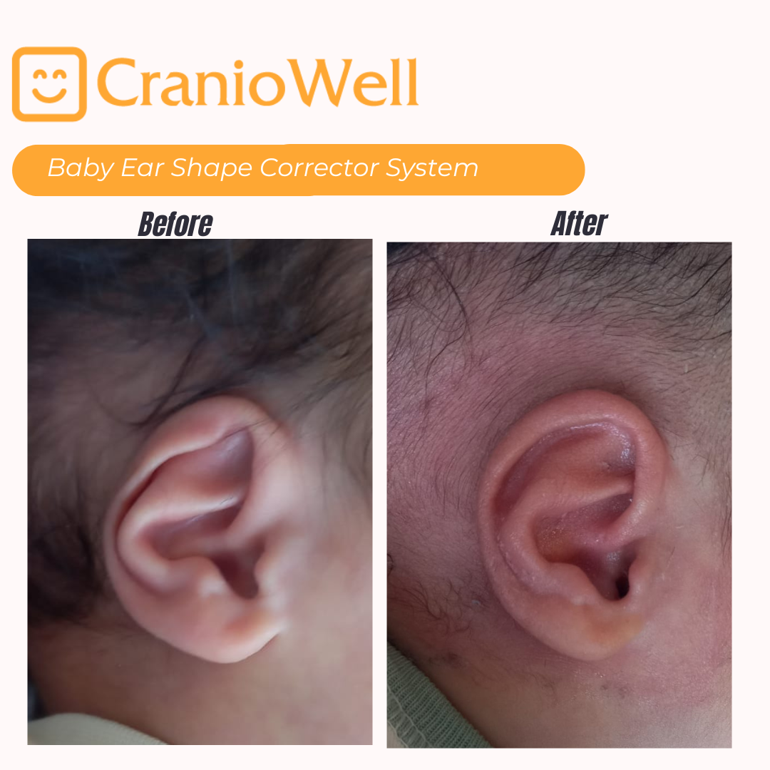
These names matter because many “folded ear” or “pointed ear” searches map onto one landmark. See also types of newborn ear deformities.

Molding acts on soft cartilage of the pinna, mainly helix, antihelix, and conchal folds. It does not rebuild missing tissue (as in microtia) and does not remove ear tags or pits. Timing and candidacy: candidate checklist and the full ear molding for infants guide.
The CranioWell baby ear corrector set is applied and followed only by treating physicians. Find the nearest one in our specialist directory. If there is no physician near you, send us a photo of your baby’s ear and your location, and we will tell you what options are available.
Shape library: prominent ears, Stahl’s ear, lop ear, cup ear, ear lidding, flat ear helix / helical rim, cryptotia, constricted ear, conchal crus, mixed ear deformity. Hub: baby ear molding.
Related: what ear molding is, do babies grow out of ear deformities?, flattening newborn ears at home.
Usually not. Molding focuses on cartilaginous folds of the upper and mid pinna, not the soft lobule.
No. Helix, antihelix, and concha are enough to describe most newborn photos to a clinician.
No. Diagrams help language; only an in-person or physician-reviewed photo assessment decides next steps.
No CranioWell physician in your country yet? You can contact us about a distribution / partnership application for your region.