Craniosynostosis in Babies: Types, Surgery & Helmet Therapy
Craniosynostosis: what it is, sagittal, coronal, metopic & lambdoid types, diagnosis, surgery options, and when helmet therapy is used after endoscopic repair. Parent guide by CranioWell.
Craniosynostosis is a birth condition in which one or more of the fibrous joints (sutures) between the skull bones close too early, before the brain has finished growing. Because the skull cannot expand in the normal direction, it compensates by growing in other directions — producing a distinctive head shape depending on which suture is affected.
Craniosynostosis affects approximately 1 in 2,000–2,500 live births. Most cases involve a single suture (simple craniosynostosis); a small proportion involve multiple sutures and may be part of a genetic syndrome.
What are the types of craniosynostosis?
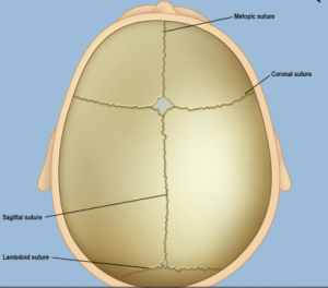
Sagittal synostosis (scaphocephaly): The most common type — 53–60% of all cases. The sagittal suture (running front to back at the top of the skull) closes early, causing the head to grow long and narrow. Related: scaphocephaly guide.
Coronal synostosis: 17–29% of cases. One or both coronal sutures (running ear to ear) close early, causing a flattened forehead on the affected side.
Metopic synostosis (trigonocephaly): 4–10% of cases. The metopic suture (running from the nose to the top of the skull) closes early, producing a triangular forehead. Related: trigonocephaly guide.
Lambdoid synostosis: The rarest type, affecting ~2% of cases. The lambdoid suture (at the back of the skull) closes early, causing flattening at the back of the head on one side — often confused with positional plagiocephaly.
Syndromic craniosynostosis: Multiple sutures involved; associated with conditions such as Apert, Crouzon, or Pfeiffer syndrome. Accounts for fewer than 5% of all craniosynostosis cases.
What are the signs of craniosynostosis?
Craniosynostosis is usually identified shortly after birth. Key signs include:
Abnormal head shape: The specific shape depends on which suture has closed.
Missing or prematurely closed fontanelle (soft spot): The anterior fontanelle may close earlier than expected or feel unusually rigid.
A raised, hard ridge along a suture: Felt along the fused suture line.
Slow or absent growth of head circumference: The head circumference curve may plateau over time.
Unlike positional plagiocephaly, craniosynostosis is caused by premature suture fusion — not sleeping position — and cannot improve on its own without treatment.
How is craniosynostosis diagnosed?
Diagnosis typically follows a physical examination in which the physician palpates the skull for fused sutures, rigid ridges, and abnormal fontanelle position. Imaging studies used to confirm the diagnosis include:
CT scan (computed tomography): The gold standard for visualising suture fusion and planning surgery. Provides 3D reconstruction of the skull.
X-ray: May show suture closure but provides less detail than CT.
MRI: Used when there is concern about intracranial pressure or brain abnormalities.
Genetic testing is recommended when syndromic craniosynostosis is suspected (multiple sutures involved or associated facial features).
What causes craniosynostosis?
In most babies, the exact cause is unknown. Known contributing factors include:
Genetic mutations: Single-gene mutations can cause syndromic craniosynostosis (e.g., FGFR2, TWIST1 mutations).
Thyroid disease in pregnancy: Mothers with thyroid disease or those treated for it during pregnancy have a higher likelihood of having a baby with craniosynostosis.
Fertility medications: Some studies report a link between clomiphene citrate use before or in early pregnancy and craniosynostosis.
Intrauterine constraint: Physical pressure on the developing skull in the womb may play a role in some cases.
What are the treatment options for craniosynostosis?
Most types of craniosynostosis require surgery. The goal of surgery is to relieve pressure on the brain, correct the skull shape, and allow normal brain growth. Surgery is typically performed in the first year of life; timing depends on which sutures are fused and whether a genetic syndrome is present.
Open cranial vault remodelling: A larger incision is made; bone segments are repositioned and reshaped. Suitable for older infants and complex cases.
Endoscopic (minimally invasive) surgery: A narrow endoscope is used to open the fused suture through small incisions. Lower risk of complications, shorter hospital stay, and less blood loss. Must be performed in the first few months of life.
Is helmet therapy used after craniosynostosis surgery?
After endoscopic craniosynostosis surgery, a custom cranial helmet (orthosis) is often prescribed to guide skull growth as the brain expands. This is different from helmet therapy used for positional plagiocephaly.
Post-endoscopic helmet: Worn for several months after minimally invasive surgery to support optimal skull reshaping. Typically started within weeks of surgery.
Not indicated after open surgery: After open vault remodelling, the skull bones are repositioned directly; a helmet is generally not required.
CranioWell HKM Pro: The HKM Pro cranial helmet is a Turkish-manufactured, patent-registered, medical device (ÜTS & EUDAMED registered) produced with Hacettepe Technopark R&D. It is applied exclusively at CranioWell Orthotics and Prosthetics Centres using 3D scanning. Related: helmet therapy guide, cranial helmet therapy.
Craniosynostosis surgery: what do parents need to know?
Surgery for craniosynostosis is performed by a neurosurgeon and/or craniofacial surgeon. Key points for parents:
Timing matters: Early surgery (before 12 months) generally produces the best outcomes because the skull is still highly malleable.
Regular follow-up is essential: After surgery, children need ongoing monitoring to ensure proper brain and skull development.
Developmental support: Some children with craniosynostosis may benefit from early intervention services; developmental delay is more common in syndromic cases.
Mild cases: Very mild single-suture craniosynostosis may not require surgery if brain pressure is absent and the head shape deformity is minimal.
Craniosynostosis occurs in approximately 1 in 2,000–2,500 live births. Single-suture synostosis is the most common form.
What is the most common type of craniosynostosis?
Sagittal synostosis accounts for 53–60% of all craniosynostosis cases. It is followed by coronal synostosis (17–29%), metopic synostosis (4–10%), and lambdoid synostosis (~2%).
Is craniosynostosis hereditary?
Most non-syndromic craniosynostosis cases are not directly inherited. Syndromic forms (affecting fewer than 5% of cases) often involve dominant gene mutations and may be inherited from a parent or arise as a new (de novo) mutation.
Can craniosynostosis be mistaken for positional plagiocephaly?
Yes — this is one of the most important distinctions. Lambdoid synostosis can mimic positional plagiocephaly. Key differences: in craniosynostosis, the ear is displaced forward on the affected side; there may be a palpable ridge; and the deformity does not improve with repositioning. A specialist evaluation and CT scan confirm the diagnosis.
Does craniosynostosis affect brain development?
In most cases of single-suture craniosynostosis treated with early surgery, cognitive and developmental outcomes are normal. In syndromic or multi-suture cases, or when diagnosis and treatment are delayed, there is a higher risk of elevated intracranial pressure, which can affect brain development.