Why Are Infant Head Shape Differences Increasing?
Since the 1990s, infant head shape differences have become more common. That rise coincides with back-sleeping recommendations that reduce sudden infant death syndrome (SIDS). Back sleeping remains essential for safety, but long periods in one position—and heavy use of modern carriers—can set the stage for deformational shape differences.
Factors That Contribute to Head Shape Differences
Intrinsic (congenital) factors
Craniosynostosis, certain genetic and metabolic syndromes, and hydrocephalus can directly affect head shape. These conditions need surgical or specialized medical follow-up and are outside the treatment scope of CranioWell HKM Pro.
Extrinsic (acquired) factors
At CranioWell we focus on deformational differences that appear after birth. Torticollis, prolonged back lying, excessive car-seat and stroller use, insufficient tummy time, and one-sided feeding habits all play roles. Premature infants have softer bones and higher risk.
How Torticollis Affects Asymmetry
Torticollis is one of the main triggers of cranial asymmetry. Weak or shortened neck muscles keep the baby turning one way. Prolonged contact on that side quickly flattens the skull. Early physiotherapy, correct positioning, and—when needed—helmet therapy work together.
Most Common Head Shape Patterns
Deformational plagiocephaly
Characterized by one-sided flattening, forehead/face asymmetry, and ear shift. Mild cases may be monitored with positioning and exercises; moderate to severe cases may be offered a custom cranial orthosis (CranioWell HKM Pro).
Symmetric brachycephaly
The head is wider than typical side to side. Marked brachycephaly can even make rolling and turning harder. Custom helmet therapy aims to normalize that width in a controlled way.
Asymmetric brachycephaly
Combines asymmetry with abnormal width. Clear frontal and vertex differences are common and often need active treatment.
Deformational scaphocephaly
A long, narrow head. Less common, but can appear in premature infants after long side-lying periods in intensive care.
Deformational differences come from external pressure and usually respond to repositioning and helmet therapy. Craniosynostosis is early suture fusion and may require surgery. A raised hard ridge along a suture, lack of bossing opposite a flat spot, and the direction of ear shift help differentiate. Suspicious cases are referred to appropriate specialists.
Positioning, Exercise, and Early Care
Correct positioning especially in the first 3 months can make a real difference. Supervised tummy time while awake, early physiotherapy for torticollis, alternating feeding sides, and limiting long carrier use are core steps. When these are not enough, CranioWell HKM Pro helmet therapy is considered at CranioWell Orthotics & Prosthetics Centers.
CranioWell HKM Pro – Custom Cranial Helmet Therapy
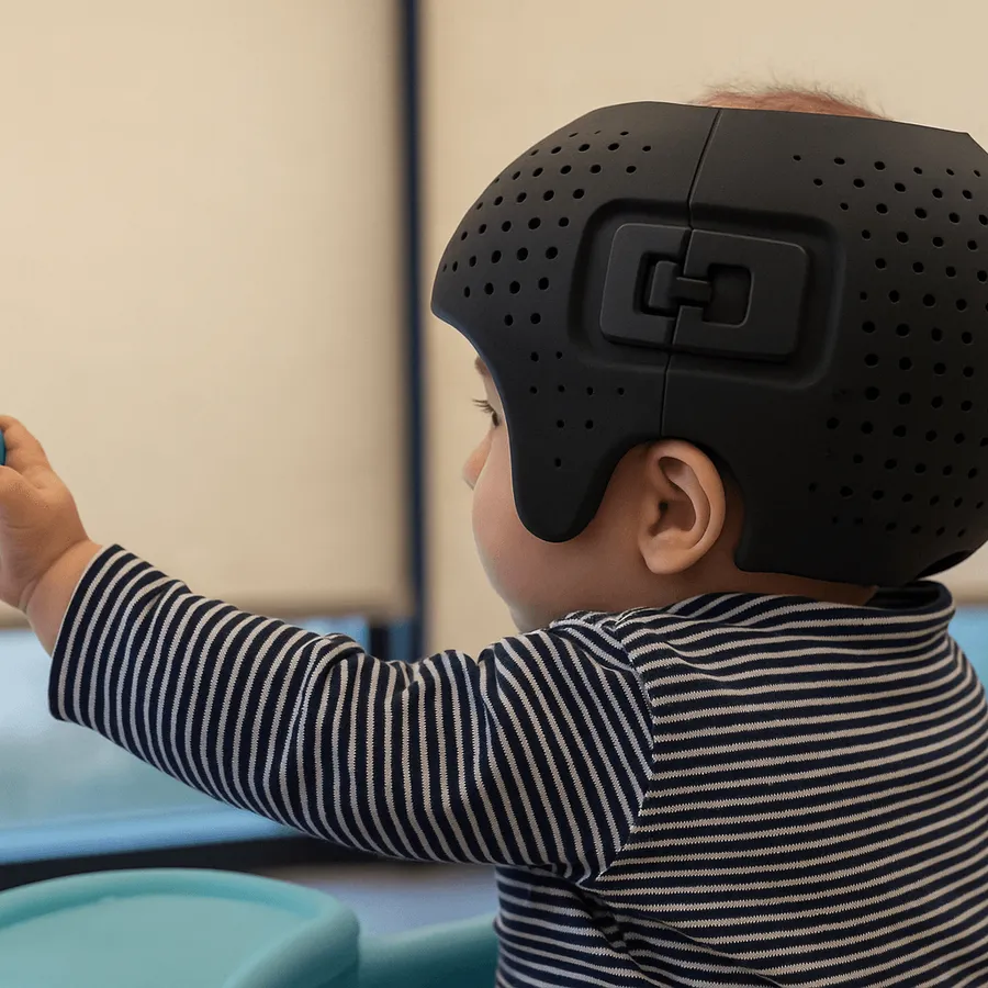
CranioWell HKM Pro helmets are medical orthoses that guide skull growth to improve symmetry and proportions. Every baby’s head is unique, so HKM Pro is not a mass-produced helmet but a custom treatment device.
Advantages of HKM Pro
- Digitally modeled for each baby
- Lightweight, breathable medical materials
- Space left over flatter areas while controlling growth where the head is already full
- Inner surface adjustable during treatment
- Closer fit and comfort than off-the-shelf designs
LED 3D Scanner – CranioWell Measurement Technology
At CranioWell, head measurement uses a high-precision LED optical scanner. It uses no radiation, is designed with infant eye and skin safety in mind, and captures full surface data in seconds without contact. It can be used even for premature and sensitive babies.
The CranioWell LED scanner is a modern optical system based on light reflection and high-resolution cameras, distinct from laser-based STARscanner workflows.
How Does Treatment Proceed?
1. Evaluation
At the first visit, history is taken, head shape is assessed, and parameters such as CVA, diagonal difference, cephalic ratio, and neck range of motion are calculated. Torticollis is specifically examined.
2. LED scanning and 3D modeling
The LED 3D scanner models the head in seconds. Data drive custom HKM Pro design.
3. Helmet fabrication
CNC processing from the digital model creates an inner surface matched to the baby. Pressure and relief zones follow the treatment plan.
4. Fitting and family education
At fitting, comfort at the forehead, ears, and occiput is checked. Families receive guidance on daily wear time, skin care, and follow-up visits. Application occurs only at CranioWell Orthotics & Prosthetics Centers.
5. Regular follow-up
Periodic visits include new measurements, inner-surface adjustments for growth, and progress review. Average duration is about 3–6 months; earlier starts within the growth window generally respond more readily.
Clinical Observations With HKM Pro
In babies treated with CranioWell HKM Pro, clinicians often observe improvement in posterior flattening, better ear alignment, reduced frontal asymmetry, and cephalic ratio moving toward a typical range. As head form improves, neck and trunk control, rolling, sitting, visual tracking, and balance can also be supported.
Summary: Structured Care With CranioWell HKM Pro
CranioWell HKM Pro is a custom, non-invasive option for deformational head shape differences, grounded in clinical practice and 3D measurement. LED-based scanning supports precise follow-up. Early care can help both appearance and function. If you are concerned about your baby’s head shape, contact a CranioWell Orthotics & Prosthetics Center for evaluation and information about HKM Pro helmet therapy.