
Head Shape
Craniosynostosis
Craniosynostosis types, frequency, risk factors, diagnosis, and treatment—including when post-surgical cranial helmet therapy may support skull shaping.
Head Shape
Craniosynostosis types, frequency, risk factors, diagnosis, and treatment—including when post-surgical cranial helmet therapy may support skull shaping.
Craniosynostosis is a birth defect in which one or more skull bones join too early—before the baby’s brain has finished growing. As the brain expands, the skull may become misshapen.
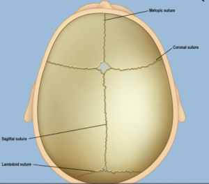
In a typical infant skull, the spaces between bones are filled with flexible tissue called sutures. These sutures allow the skull to grow as the brain grows. Around age two, the skull bones begin to fuse as sutures turn to bone—“closing.” In craniosynostosis, one or more sutures close too early. This can limit or slow the baby’s brain growth.
Growth stops only in the area of the prematurely closed suture. Other open regions continue to grow. The brain may reach a normal size while the skull takes an abnormal shape. Sometimes several sutures close early; then the brain may not have enough room, and pressure inside the skull can rise.
Types depend on which sutures fuse early.
The sagittal suture runs along the top of the head from the soft spot in front to the back. When it closes too early, the head becomes long and narrow (scaphocephaly). It is the most common type of craniosynostosis.
The right and left coronal sutures run from each ear up to the sagittal suture. When one closes early, the forehead on that side may flatten (anterior plagiocephaly). The eye socket on that side may sit higher and the nose may pull toward that side. This is the second most common type. Read more about plagiocephaly.
This type occurs when both coronal sutures close early. The head becomes wide and short (brachycephaly).
The lambdoid suture runs across the back of the head. Early closure can flatten the back of the head (posterior plagiocephaly). It is among the rarer types.
The metopic suture runs from the nose up to the sagittal suture. Early closure can give a triangular head shape—narrow in front and wider behind (trigonocephaly). It is also among the rarer types. Read more about metopic synostosis.
Most problems a baby may face depend on:
If untreated in some cases, excess pressure inside the skull can lead to problems such as blindness, seizures, or brain injury.
Researchers estimate that about 1 in every 2,500 babies in the United States is born with craniosynostosis. Clear nationwide figures for Turkey are not yet available from published studies.
In most babies, the causes of craniosynostosis are unknown. Some infants have craniosynostosis because of gene changes. In some cases, abnormality in a single gene causes a genetic syndrome. In most cases, craniosynostosis is thought to result from a combination of genes and environmental factors—such as substances the mother contacts, eats, or drinks, or certain medicines used during pregnancy.
Recent reports have linked higher chance of having a baby with craniosynostosis to:
Craniosynostosis is often diagnosed soon after birth. Sometimes diagnosis comes later.
Usually the first sign is an abnormally shaped skull. Other signs may include:

A physician may identify craniosynostosis during physical examination by feeling along the sutures for hard ridges and unusual soft spots, and by assessing facial shape.
If craniosynostosis is suspected, one or more tests are usually ordered. Special imaging such as CT (CAT) scan can show skull and brain detail, whether specific sutures are closed, and how the brain is growing.
Many types of craniosynostosis require surgery to relieve pressure on the brain, correct the synostosis, and allow proper brain growth. When needed, surgery is often done in the first year of life. Timing depends on which sutures closed and whether a genetic syndrome is present.
Surgery may be open or endoscopic (minimally invasive). Open surgery generally carries a higher risk of complications; endoscopic approaches have lower complication risk in suitable patients. After endoscopic surgery, helmet therapy may be recommended to support ongoing skull shaping. Helmets are fitted only at CranioWell Orthotics & Prosthetics Centers.
Mild cases
Some infants with very mild craniosynostosis may not need surgery. As the child grows and hair grows in, the skull shape may become less noticeable.
Every baby with craniosynostosis is different; severity ranges from mild to severe. Most are generally healthy. Some children have developmental delays or intellectual disability because synostosis limited brain growth or because a genetic syndrome affects both the skull and brain function.
Infants with craniosynostosis need regular follow-up to confirm that brain and skull are developing properly. Early intervention can help many families.
Some children struggle with self-esteem because of appearance differences. Parent support groups for craniofacial birth differences can also be helpful.